The 3D Mammography Exam
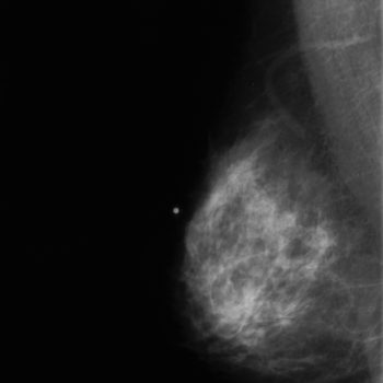
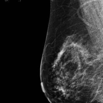
3D mammography, also known as tomosynthesis, is an advanced technology that shows is helping in the fight against breast cancer. It has specially designed digital detectors to capture multiple segments or ‘slices’ of the breast at different angles and then reconstructs them into a three-dimensional image that can be displayed in high resolution.
A 3D mammogram is very much like having a digital mammogram. The process is performed at the same time as a traditional 2D mammogram, on the same scanner with no noticeable difference to the patient. While the breast is compressed, a second set of images is obtained to create a 3D image of the breast so the radiologist can evaluate the breast tissue one “slice” at a time.
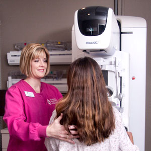
During your mammography at the Women’s Imaging Center, a specially qualified radiologic technologist will position you to image your breast. The breast is placed on a special platform and compressed with a paddle (often made of clear Plexiglas or other plastic). Our staff is caring and will ensure you are comfortable throughout the process.
The Women’s Imaging Center is accredited by the American College of Radiology and has been designated as a Breast Imaging Center of Excellence. To be designated as a breast center of excellence, breast imaging centers are fully accredited in mammography, breast ultrasound, breast ultrasound-guided breast biopsy and stereotactic breast biopsy.
To schedule your 3D mammogram with Women’s Imaging Center, please call us at (863) 688-2334.
Breast compression for your 3D mammogram is necessary in order to:
- Even out the breast thickness so that all of the tissue can be visualized.
- Spread out the tissue so that small abnormalities won’t be obscured by overlying breast tissue.
- Allow the use of a lower X-ray dose since a thinner amount of breast tissue is being imaged.
- Hold the breast still in order to eliminate blurring of the image caused by motion.
- Reduce X-ray scatter to increase the sharpness of the picture.
The technologist will be in the examination room at all times. You will be asked to change positions slightly between images. The routine views are a top-to-bottom view and a side view. The process is repeated for the other breast. The screening mammography process should take about 15 minutes. The images are immediately available for the radiologist to review.
The Women’s Imaging Center is located in Lakeland, Florida. We serve patients from Central Florida including Plant City, Winter Haven, Auburndale, Kissimmee, and more. Please call us at (863) 688-2334 to schedule your screening or diagnostic mammogram.
Frequently Asked Questions About 3D Mammography at the Women’s Imaging Center:
What is 3D mammography?
3D mammography is a new technology that shows great promise in the fight against breast cancer. It uses computers and specially designed digital detectors to produce an image that can be displayed on a high-resolution computer monitor. From a patient’s point of view, having a 3D mammogram is very much like having a conventional film mammogram.
Why is 3D mammography important?
Mammography is used to aid in the diagnosis of breast diseases in women. Mammography plays a central part in the early detection of breast cancers. Successful treatment of breast cancer often is linked to early diagnosis. Studies have shown that 3D mammography can be more effective in women with dense breasts and women younger than 50.


